Arteries In Neck : Anatomy Of The Neck Vessels. The carotid arteries are two large blood vessels that supply oxygenated blood to the large, front part of the brain. Stroke deprives your brain of oxygen. Carotid arteries are major arteries that carry blood from your heart to your brain. A buildup of plaque can narrow or block your carotid arteries. Carotid arteries are located in the anterior of the neck, on either side.
Once these complications occur patients develop certain symptoms and signs such as. However, neck arteries can work just as fine, even though they are partially blocked. The left and right common carotid arteries ascend up the neck, lateral to the trachea and the oesophagus. These blood vessels can have abnormal shapes, sizes or paths through the neck and head. Your doctor may then test your physical and mental capabilities such as strength, memory and speech.
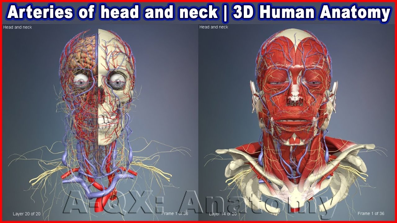
Arteries of head and neck | 3D Human Anatomy | Organs ... from i.ytimg.com In case blood clots dislodge, they travel through brain vessels and eventually end up clogging vessels of smaller diameter than they are. The vein is the most lateral structure within the carotid sheath, followed by the nerve and then the artery, which is the most medial structure. With a cervical artery dissection, the neck pain is unusual, persistent, and often accompanied by a severe headache, says dr. The left and right common carotid arteries ascend up the neck, lateral to the trachea and the oesophagus. These blood vessels can have abnormal shapes, sizes or paths through the neck and head. What are the arteries of the chest, neck and brain? Plaques in the neck arteries increase the risk of blood clots. Without this blood flow, your brain cells would.
At the root of the neck the right internal jugular vein is placed at a little distance from the common carotid artery, and crosses the first part of the subclavian artery, while the left internal jugular vein usually overlaps the common carotid artery.
Carotid artery stenosis is a major risk factor for stroke. The carotid arteries can be felt on each side of the lower neck, immediately below the angle of the jaw. They supply oxygen to the parts of the brain that control our movements and our ability to think, speak and experience our senses of touch, taste, sight, sound and feel. Carotid artery blockage, also known as carotid artery stenosis, is the constriction of the inner surface of the carotid artery due to buildup of plaque. The carotid arteries are two large blood vessels that supply oxygenated blood to the large, front part of the brain. The carotid artery brings needed blood to your brain and face. This is a cause of transient ischemic attack or a stroke. Two major arteries of the thorax that lie beneath the clavicles.the vertebral arteries ascend through the neck inside the transverse foramina of the cervical vertebrae, all the way to the brain.through their course, they give off several meningeal, muscular and spinal branches for the nearby structures. When your doctor puts their hands on your neck to detect. You have two carotid arteries, one on each side of your neck. The blockage increases your risk of stroke, a medical emergency that occurs when the blood supply to the brain is interrupted or seriously reduced. The vertebral arteries stem from the subclavian arteries; Once these complications occur patients develop certain symptoms and signs such as.
This is called carotid artery disease, which increases your risk of stroke. The blockage increases your risk of stroke, a medical emergency that occurs when the blood supply to the brain is interrupted or seriously reduced. A buildup of plaque can narrow or block your carotid arteries. Carotid arteries are located in the anterior of the neck, on either side. Once these complications occur patients develop certain symptoms and signs such as.
Major blood vessels of the neck - YouTube from i.ytimg.com Carotid arteries are located in the anterior of the neck, on either side. In simpler terms, a carotid artery blockage is a result of the blood vessels in the neck getting blocked due to the buildup of plaque on the arterial walls. When your doctor puts their hands on your neck to detect. The left and right common carotid arteries ascend up the neck, lateral to the trachea and the oesophagus. A buildup of plaque can narrow or block your carotid arteries. At the level of the superior margin of the thyroid cartilage (c4), the carotid arteries split into the external and internal carotid arteries. Carotid artery disease occurs when fatty deposits (plaques) clog the blood vessels that deliver blood to your brain and head (carotid arteries). However, neck arteries can work just as fine, even though they are partially blocked.
These blood vessels can have abnormal shapes, sizes or paths through the neck and head.
The pain may be sudden and severe—people often describe it as a throbbing pain. A new classification system divides the internal carotid artery into four parts; A vertebral artery tear may feel like something sharp is stuck in the base of your skull. They do not give off any branches in the neck. There are two carotid arteries (one on each side of the neck) that supply blood to the brain. However, neck arteries can work just as fine, even though they are partially blocked. Related posts of arteries in the neck picture veins and arteries of the neck. The left and right common carotid arteries ascend up the neck, lateral to the trachea and the oesophagus. The internal carotid artery is one of two branches of the common carotid artery. Blood is carried to the brain through blood vessels called arteries. Veins and arteries of the neck 9 photos of the veins and arteries of the neck activate javascript arteries in the neck diagram, common carotid artery branches, external carotid artery function, how many carotid arteries, left common carotid artery function, the left common carotid artery supplies blood to the. At the root of the neck the right internal jugular vein is placed at a little distance from the common carotid artery, and crosses the first part of the subclavian artery, while the left internal jugular vein usually overlaps the common carotid artery. When your doctor puts their hands on your neck to detect.
They are the carotid arteries, and they carry blood to the brain. The exam generally includes listening for a swooshing sound (bruit) over the carotid artery in your neck, a sound that's characteristic of a narrowed artery. However, pain from carotidynia typically only occurs on one side. The arteries in the chest, neck and brain are the most frequent arteries found to be abnormal in phace syndrome. Arteria carotis interna) is located in the inner side of the neck in contrast to the external carotid artery.
23 The face and superficial neck | Pocket Dentistry from pocketdentistry.com You have one of these arteries on each side of your neck. The carotid arteries are major blood vessels in the neck that supply blood to the brain, neck, and face. With a cervical artery dissection, the neck pain is unusual, persistent, and often accompanied by a severe headache, says dr. The arteries in neck that supply blood to the brain are called carotid arteries. This is called carotid artery disease, which increases your risk of stroke. Without this blood flow, your brain cells would. The blockage increases your risk of stroke, a medical emergency that occurs when the blood supply to the brain is interrupted or seriously reduced. In the neck, the carotid sheath (fibrous connective tissue) covers the common carotid artery, vagus nerve, and internal jugular vein.
The neck pain from a carotid artery tear often spreads along the side of the neck and up toward the outer corner of the eye.
There are two large arteries in the neck, one on each side. Blood is carried to the brain through blood vessels called arteries. The carotid arteries are major blood vessels in the neck that supply blood to the brain, neck, and face. Plaques in the neck arteries increase the risk of blood clots. In the neck, the carotid sheath (fibrous connective tissue) covers the common carotid artery, vagus nerve, and internal jugular vein. At the level of the superior margin of the thyroid cartilage (c4), the carotid arteries split into the external and internal carotid arteries. This is a cause of transient ischemic attack or a stroke. The neck pain from a carotid artery tear often spreads along the side of the neck and up toward the outer corner of the eye. At the root of the neck the right internal jugular vein is placed at a little distance from the common carotid artery, and crosses the first part of the subclavian artery, while the left internal jugular vein usually overlaps the common carotid artery. The blockage increases your risk of stroke, a medical emergency that occurs when the blood supply to the brain is interrupted or seriously reduced. There are two carotid arteries, one on the right and one on the left. Just like other arteries in the body, neck arteries are also susceptible to blockages. The plaque buildup is made of fat, cholesterol, cellular waste, calcium, proteins and inflammatory cells.